Read Cytohistology of Focal Liver Lesions (Cytohistology of Small Tissue Samples) - Aileen Wee | PDF
Related searches:
3943 4668 1899 3475 406 988 1983 4161 4076 1670 3780 3885 967 1999 2970 2954 2345 4780 449 367 3518 4894 3044 2218 1042 3906 1099 248 3060 4655 4445 4224 2490 220 446 3135 1623 2791
Benign focal liver lesion contrast agent ct liver magnetic resonance malignant focal liver lesion ultrasound focal liver lesions (fll) are common in the general population and are frequently found during ultrasound examination either incidentally, in healthy subjects, in symptomatic patients and in patients with oncological history, during.
The calculated prevalence of benign focal liver lesions shows that on the fortuitous discovery of space-occupying lesions of the liver, first consideration should be given to focal fatty sparing, simple hepatic cysts and hemangiomas. The finding of a fnh or an adenoma is rarely a random discovery.
The main liver application of ceus is for focal liver lesions. Focal liver lesions are usually detected incidentally during an abdominal ultrasound examination, during first evaluation or follow-up for a primary neoplasm, or during surveillance in chronic liver diseases and cirrhosis.
The discovery of focal liver lesions (flls) is a frequent clinical situation in daily abdominal imaging practice. Contrast material–enhanced ultrasonography (us) has demonstrated its efficacy in the characterization of flls with a diagnostic performance similar to that of computed tomography (ct) and magnetic resonance (mr) imaging (1–10), without the use of ionizing.
Diagnostic approaches and management of focal liver lesions (flls). Flls are solid or cystic masses or areas of tissue that are identified as an abnormal part of the liver. The term “lesion” rather than “mass” was chosen because “lesion” is a term that has a wider application, including solid and cystic masses.
Liver-specific contrast agents, mri is able to not only provide morphological and vascular information of the focal liver lesions, but also functional information on the capacity of the lesion to uptake the liver-specific contrast agent. The main drawback of the technique is the requirement for state-of-the-art.
Focal liver lesions are abnormal solid or liquid masses differentiated from normal liver through cross-sectional imaging 1, 2 usually detected incidentally via imaging due to unrelated symptoms typically clinically silent but large lesions may be associated with right upper quadrant abdominal pain.
The authors discuss the commoner focal liver lesions encountered and the methods available for further investigation #### learning points a 31 year old anglo-indian man presented to his general practitioner for a discussion of cardiovascular risk factors because of a strong family history of ischaemic heart disease.
Your liver is an important organ, and disease can prevent it from working the way it should to keep you healthy. Learn about the symptoms and stages of liv your liver is an important organ that performs a wide range of functions, including.
The causes of hypodensity liver lesions are many and they could include benign liver cysts that have no symptoms or malignant tumors which are usually associated with certain symptoms. Sometimes a part of the liver tissue may become hypodense as compared to the nearby tissue due to focal fatty changes or due to primary or secondary tumors.
Nonalcoholic fatty liver disease is a common condition that can lead to serious problems. High blood what can we help you find? enter search terms and tap the search button.
A hypodense liver lesion or hypodensity liver is a deformity in the liver tissue that appears less dense than the surrounding tissue in radiological scans such as computed tomography (ct) scans or magnetic resonance imaging (mri). The appearance of these lesions in the radiological tests does not improve with the injection of intravenous.
The general definition of a lesion is an area of atypical tissue, according to the national cancer institute. Lesions can be either cancerous (malignant) or benign, meaning not cancerous.
Liver lesions are abnormal clumps of cells in your liver, and they are very common.
Out of various pathologies that affect the liver, focal liver lesion form an important group. Triphasic computed tomography is the imaging modality most often used to evaluate focal liver lesions.
The term lesion is broad and includes any wound, sore, ulcer, tumour or other tissue damage of an organ. A focal lesion in the liver refers to one area of tissue damage identified on the liver that has varying significance depending on the patient's health condition and a variety of other factors.
The most common benign solid lesions of the liver represent congenital vascular lesions that contain fibrous tissue and small blood vessels that eventually grow range in size from small (1 centimeter or less) to giant, cavernous hemangiomas (10 to 20 centimeters) spontaneous rupture (bleeding) is rare.
To characterize the focal liver lesion with confidence as either needing no or only routine follow-up. To determine which ones needing further, more rigorous exploration (including biopsy). Managing some of these focal liver lesions thru primary care practice.
Cytohistology of focal liver lesions aileen wee mixed media product. Breast cytohistology with dvd-rom joan cangiarella mixed media product.
This pathway provides guidance on imaging in patients with focal liver lesions, dependent on whether the patient has risk factors for primary cancer or metastases or whether the lesion is ‘incidental’.
Liver lesions detected by a ct scan can be caused by many things, ranging from benign cysts to liver cancer, according to sutter health california pacific medical center. Many of these conditions are asymptomatic and have few or no long-term health consequences. The most common type of liver lesions are hemangiomas, reports sutter health cpmc.
Few fields of medicine have witnessed such impressive progress as the diagnosis and treatment of liver tumors. Advances in imaging technology, the development of novel contrast agents, and the introduction of optimized scanning protocols have greatly facilitated the non-invasive detection and characterization of focal liver lesions.
The usual differential diagnosis of multiple, focal lesions in liver and spleen include lymphoma, leukaemia deposits, metastasis, bacterial and fungal infection, and sarcoid. Most of these diseases give rise to non-specific focal hypoechoic lesions on sonography.
More importantly, the evaluation of liver lesions has taken on greater importance because of the increasing incidence of primary hepatic malignancies, especially hepatocellu-lar carcinoma (hcc) and cholangiocarcinoma (cca). Th erefore, a thorough and systematic approach to the management of focal liver lesions (flls) is of utmost importance.
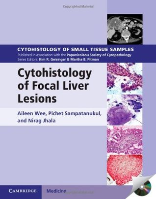
This book provides comprehensive coverage of both surgical pathology and cytopathology of focal liver lesions. Benign, pre-malignant and malignant entities are presented with key morphologic criteria, example vignettes, flowcharts, over 700 printed photomicrographs and a cd-rom offering all images in a downloadable format.
Viruses such as hepatitis c can cause liver inflammation and damage. Other liver conditions can be the result of drugs or drinking too much alcohol.
Focal nodular hyperplasia is a benign tumor growth in the liver. Focal nodular hyperplasia is the second most common benign growth in the liver after hemangioma. Most cases are asymptomatic and are only diagnosed accidentally from imaging studies, while one third of the cases are discovered due to the emergence of clinical symptoms.
Your liver is an important organ that performs a wide range of functions, including aiding digestion and removing toxins from your body. Like many of the other organs in your body, your liver is also susceptible to developing disease, which.
Benign tumor-like lesion of the liver which is considered to be the result of a hyperplastic response of the hepatocytes to the presence of a pre-existing vascular malformation.
The american college of gastroenterology has issued a new guideline on the diagnosis and management of focal liver lesions, now that widespread use of imaging has increased detection rates.
This volume provides comprehensive coverage of both surgical pathology and cytopathology of focal liver lesions. Extensively illustrated throughout, it contains key cytologic and histologic features, practical points, radiologic and morphologic pictures, flow charts, and tabulated summaries for easy comprehensive overview and quick reference.
Liver lesions are abnormal clumps of cells in your liver, and they are very common. They will be detected in as much as 30% of people over 40 who undergo imaging tests. the majority of liver lesions are benign (not harmful) and don't require treatment. But in some cases, liver lesions are malignant (cancerous) and should be treated.
Focal nodular hyperplasia (fnh) is a regenerative mass lesion of the liver and the second most common benign liver lesion (most common is a hemangioma). Many fnhs have characteristic radiographic features on multimodality imaging, but some lesions may be atypical in appearance.
Relatively common in the liver, benign (noncancerous) liver masses or lesions may incidentally be detected on imaging studies, abnormal liver function tests, or during investigation of abdominal pain. Pain often occurs in lesions greater than 5 to 6 centimeters in size; hepatic adenomas.
Focal liver lesions are usually first detected and designated as such on imaging. A focal lesion is an area or region of abnormal or altered echogenicity on ultrasound, abnormal density on computerized tomog- raphy (ct), or abnormal signal intensity on magnetic resonance imaging (mri).
Hemangiomas, focal nodular hyperplasias (fnh), and adenomas (hca) are the most commonly encountered solid benign lesions. 1 - 3 the most commonly encountered malignant lesions in noncirrhotic livers are metastases. Hepatocellular carcinoma (hcc) and intrahepatic cholangiocarcinoma (icc) occur in the setting of chronic liver disease.
Sponsored by papanicolaou society of cytopathology and cambridge university press.
Cytohistology of small tissue samples cytohistology of focal liver lesions aileen wee professor and senior consultant pathologist, department of pathology, yong loo lin school of medicine,.
In fatty liver disease, accumulation of fat inside the liver can lead to lesions on liver. Focal fatty change is more common in people who have been diagnosed with diabetes, hepatitis c, obesity and chronic liver diseases.
Cytohistology of focal liver lesions - how to report common problematic patterns - prof. Aileen wee, national university hospital, singapore, singapore 1025 - 1100 tea break 1100 - 1145 potpourri of interesting cases in cytology - prof. Sheema h hasan, aga khan university hospital, karachi, pakistan 1150 - 1235.
If a focal liver lesion is isointense to hyperintense to liver on a t1-weighted image, then it is most commonly hepatocellular in origin. The 5 most common focal hepatocellular lesions encountered in clinical practice are regenerative nodules (rn), hepatocellular carcinoma (hcc), focal nodular hyperplasia (fnh), hepatocellular adenoma (hca), and focal steatosis.
Get the facts about liver diseases, such as hepatitis, cancer, and cirrhosis. Know your risk and what you can do to prevent liver problems.
Live a healthy lifestyle! subscribe to our free newsletters to receive latest health news and alerts to your email inbox.
Post Your Comments: